| Home | | | News | | | Basics | | | People | | | History | | | Links | | | Hot Points | | | FAQ |
"-sign.![]() |
1) Pedersen, P. L., and
Carafoli, E. (1987) Ion motive ATPases. I. Ubiquity, properties, and
significance to cell function. Trends Biochem. Sci. 4:
146-150. 2) P-type ATPase Database (By Kristian B. Alexsen, Swiss Institute of Bioinformatics) 3) Kawasaki-Nishi S, Nishi T, Forgac M. (2003 ) Proton translocation driven by ATP hydrolysis in V-ATPases. FEBS Lett. 545(1): 76-85. 4) Perzov N, Padler-Karavani V, Nelson H, Nelson N. (2001) Features of V-ATPases that distinguish them from F-ATPases. FEBS Lett. 504(3): 223-8. |

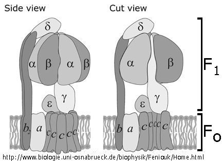
| F1 |
FO |
||
| Alpha |
3 |
a |
1 |
| Beta |
3 | b |
2 |
| Gamma |
1 |
c |
10-14(?) |
| Delta |
1 |
||
| Epsilon |
1 |
||
).
In case of hydrolysis the enzyme functions as an ATP-driven proton pump
and generates
.
o´) for this
reaction. This is a reasonable estimate, for figures from -28 to -36 kJ
mol-1
can be found in literature, the most popular being -30.6 kJ mol-1
(-7.3 kcal/mol).
o, is the total amount
of energy which is either used up or released during a chemical
reaction under standard
conditions when the chemical activities of all the reactants is
equal to 1. In case of reactions in aqueous solutions the activities
are usually substituted by concentrations (i.e. 1 M); the activity of
water itself is taken as 1. "Biological" standard Gibbs
free energy change,
o´, is a similar
parameter, but is defined at pH 7, i.e. the concentration of H+
is not 1 M, but 10-7M. It is more practical and convenient,
for most biological reactions take place at physiological pH.
o´ is not the amount of energy available
to drive other, endothermic reactions in
the cell, because the conditions in the cell are not standard
(see the definition above). The actual Gibbs energy change is
=
o'
+ 2.3 RT log [CADP
CPi (CH+ / 10-7) / CATP ],| CATP | 2 x 10⁻³ M-1 |
| CADP | 2 x 10⁻⁴ M-1 |
| CPi | 10-2 M-1 |
| CH+ | 5 x 10⁻⁸ M-1(pH
approx. 7.3) |
=
o'
+ 2.3 RT log ( CADP
CPi CH+ / CATP )
= -30 - 19.6 = - 49.6 kJ mol-1
upon the change in the
concentration
of one reactant (x axis),
assuming that the concentrations of other reactants are kept constant
at values used in the example above (red dots indicate the
calculated in this example). 
![]() |
1) Nicholls, D. G. and S.
J. Ferguson. Bioenergetics 2,
London:Academic Press, 1992. 2) Any edition of "Physical Chemistry" by P. Atkins |
composed of two
components: the chemical and the
electrical one. The more protons are on one side of a membane relative
to
the other, the higher is the driving force for a proton to cross the
membrane. As proton is a charged particle, it's movement is also
influenced by electrical field: transmembrane electrical potential
difference will drive protons from positively charged side to
the negatively charged one.
is measured in Joules per mole (J mol-1) and is
defined as:
=
-F
+ 2.3
RT (pHP - pHN),
is the
transmembrane electrical potential difference in
volts. The value of
tells, how much energy is required (or is released, depending on the
direction of the transmembrane proton flow) to move 1 mol of protons
across the membrane.
, but protonmotive force (pmf):
/
F =
-
2.3
RT/F (pHP - pHN)
)
is:
- 59 (pHP
- pHN)
between 11.6 and
19.3 kJ mol-1)
![]() |
1) Nicholls, D. G. and S.
J. Ferguson. Bioenergetics 2,
London:Academic Press, 1992. 2) A Lecture on Electrochemical potential by Prof. A.R. Crofts 3)Cramer, W.A. and D.B. Knaff. Energy Transduction in Biological Membranes: A Textbook of Bioenergetics, Springer-Verlag New York/Berlin/London |
![]() |
1) W. Junge, H. Lill, and
S. Engelbrecht. (1997) ATP synthase:
an
electrochemical transducer with rotatory mechanics. Trends Biochem.Sci. 22
(11):420-423, . 2) H. Wang and G. Oster. (1998) Energy transduction in the F1 motor of ATP synthase. Nature 396 (6708):279-282. 3) Weber, J., and Senior, A. E. (2003) ATP synthesis driven by proton transport in F1FO-ATP synthase. FEBS Lett. 545(1): 61-70. 4) Nice movies at http://nature.berkeley.edu/~hongwang/Project/ATP_synthase/ |

|
The answer is three. Not five.
However, the total amount of the nucleotide-binding
sites is six, three of them being non-catalytic. Each
site is located on the interface between subunits Alpha and Beta.
Larger part of each catalytic site is composed from aminoacid residues
of the
respective Beta-subunit, while each non- catalytic site is situated
mostly on the respective Alpha subunit. The role of the non-catalytic
sites is obscure, they are not necessary for the catalysis. One
possibility is that they facilitate the enzyme assembly in the cell.
There is also evidence that the
Epsilon subunit binds adenine nucleotides.
|
![]() |
![]() |
1) C. Etzold, G.
Deckers-Hebestreit, and K. Altendorf. (1997) Turnover number of Escherichia coli FOF1- ATP synthase for ATP synthesis in
membrane vesicles. Eur.J.Biochem.
243 (1-2):336-343. 2) R. L. Cross, C. Grubmeyer, and H. S. Penefsky. (1982) Mechanism of ATP hydrolysis by beef heart mitochondrial ATPase. Rate enhancements resulting from cooperative interactions between multiple catalytic sites. J.Biol.Chem. 257:12101-12105. 3) U. Junesch and P. Gräber. (1985) The rate of ATP synthesis as a function of Delta pH in normal and dithiothreitol-modified chloroplasts. Biochim.Biophys.Acta 809:429-434. |
| Constructed and maintained by Boris A. Feniouk |
Summer 2002 |